The respiratory system is primarily involved in the exchange of gases (carbon dioxide and oxygen) between the atmosphere and the blood that provides oxygen to the cells and removes carbon dioxide produced by aerobic respiration. It is also involved in other important life functions such as pH regulation, thermoregulation, and defense against pathogens. A series of structures allow for air to be conducted to the lungs by bulk flow. In the lungs, gas exchange occurs by diffusion. The conducting zone includes the structures that carry air from the outside to the lungs and the respiratory zone is the site of gas exchange in the lungs.
Humans need a continuous supply of Oxygen for cellular respiration & they must get rid of excess carbon dioxide, the poisonous waste product of this process.
Gas exchange supports this cellular respiration by constantly supplying oxygen & removing carbon dioxideOxygen required for respiration is obtained from Earth’s atmospheric air, which is 21% oxygen.This oxygen in the air is exchanged in the body by respiratory surface provided by the Alveoli in the lungs.
What are causes of asphyxia?
Examples of injuries or illnesses that can cause asphyxiation can include:
- Collapsed lung
- Inhalation of toxic fumes (like carbon monoxide)
- Whooping cough
- Diphtheria (bacterial infection)
- Croup
- Heart failure
- Swollen veins in the head or neck
- Paralysis
Symptoms of asphyxia at the time of birth may include:
- Not breathing or very weak breathing
- Skin color that is bluish, gray, or lighter than normal
- Low heart rate
- Poor muscle tone
- Weak reflexes
- Too much acid in the blood (acidosis)
- Amniotic fluid stained with meconium (first stool)
- Seizures
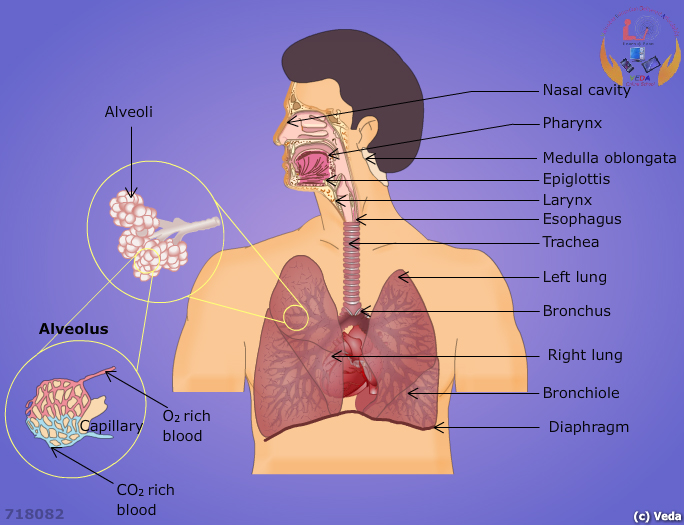
The organs of the respiratory system can be divided into two groups. The upper respiratory tract consists of the nose, nasal cavity and pharynx. The lower respiratory tract includes the larynx, trachea, bronchial tree, and lungs.
Air enters the respiratory tract through the nose and travels through the nasal cavity where it is filtered and warmed by mucous membranes and nasal hairs (also called vibrissae). Olfactory receptors are also present on the walls of the nasal cavity. The humidified air then reaches the pharynx, which is located behind the nasal cavity and at the back of the mouth. The pharynx is common to both the digestive and respiratory systems and functions as a pathway for air traveling to the lungs and food entering the esophagus. The epiglottis is an elastic cartilaginous flap that closes to keep food out of the respiratory tract. During swallowing, the larynx rises and the epiglottis covers the opening into the larynx. Two vocal cords are present in the larynx that vibrates when air passes between them.
The trachea or windpipe is a flexible tube that extends from the larynx into the thoracic cavity where it divides into the right and left primary bronchi. Cartilaginous rings are open posteriorly and function to maintain the rigidity of the trachea.
Within the lungs, the bronchi branch further into smaller structures called bronchioles. Bronchioles divide further until they end in small grape-like structures called alveoli, where gas exchange occurs. Alveoli are therefore considered the functional unit of the lungs. Surfactant is a detergent-like substance that prevents the alveolus from collapsing by lowering the surface tension. A web of blood capillaries surrounds each alveolus to carry oxygen and carbon dioxide. The extensive branching and small size of the alveoli mean that there is a large surface area for gas exchange. The right lung is divided into three lobes whereas the left lung has two lobes.
There are two types of cells that form the epithelial tissues of alveoli: Type I and Type II. Alveolar cells are also known as pneumocytes. Type I alveolar cells are the thin squamous epithelial cells that line the alveolus and form the site of gaseous exchange between the alveolus and blood. Type II alveolar cells are interspersed among the type I cells and they release the alveolar fluid and surfactant. Alveolar macrophages are found in alveoli and function to engulf foreign particles including bacteria.
A layer of membrane called the visceral pleura is attached to each lung and folds over to form the parietal pleura that attaches to the inner wall of the thoracic cavity. The space between these membranes is called the pleural cavity and it contains pleural fluid. This fluid functions to reduce friction and to assist lung expansion.
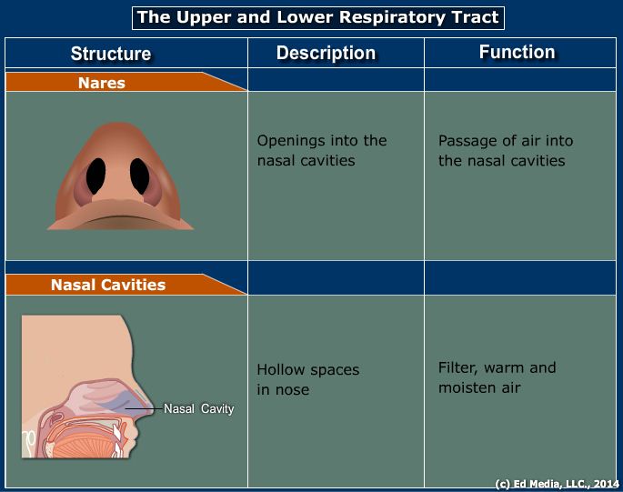
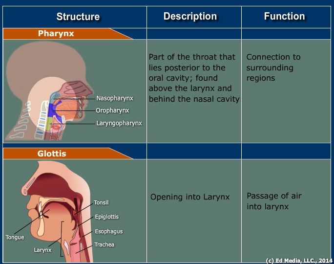
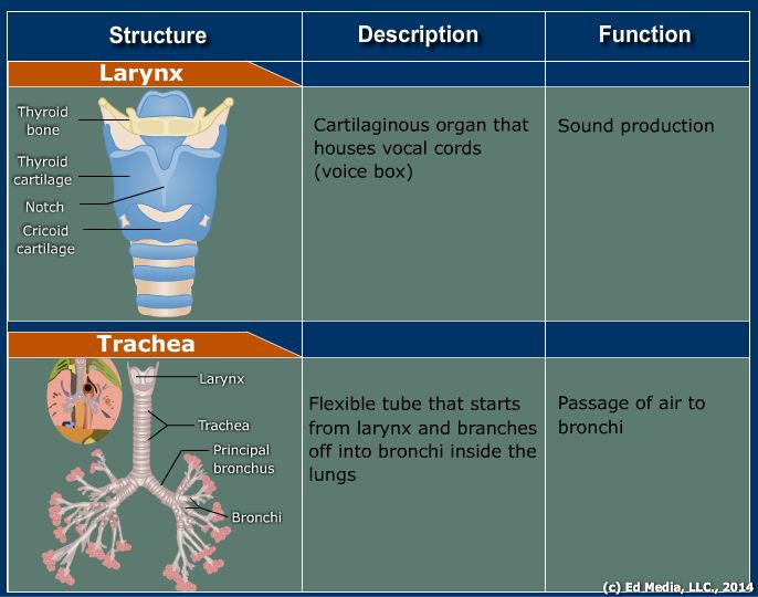
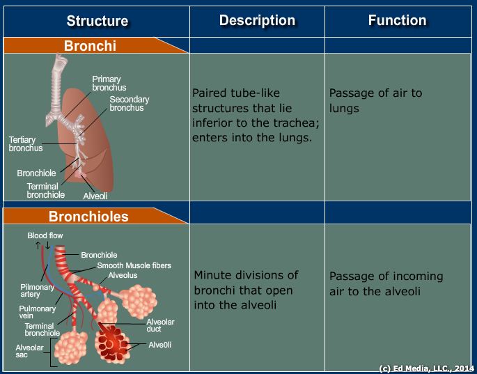
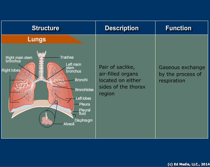
The following media explains about the Respiratory Tract:
