Introduction


The anatomical system, which enables movement, is termed the muscular system. Muscles are the contractile tissues of the body derived from embryonic germ cells. The muscular system is controlled by the nervous system in vertebrates and few muscles work autonomously. The cells making up the muscular system comprise muscle fibers. The functions of muscles include posture, balance, strength, movement and heart contraction.
Smooth musclesSmooth muscle is involuntary under the control of the autonomic nervous system. The tissue is called smooth because the arrangement of the myofibrils is homogenous and not striated. Smooth muscle tissue consists of long spindle-shaped cells with a single nucleus and is located on the walls of hollow internal organs as they help contraction, especially in the digestive tract, bladder, uterus, blood vessel walls, and the respiratory tract. Compared to other muscle types, smooth muscles contract slowly, do not fatigue easily and have sustained and prolonged contractions. Tonus refers to a constant state of contraction as observed in blood vessels.
Cardiac muscleCardiac muscle is only found in the heart and is comprised of cells that mostly contain a single nucleus. It is involuntary under the control of the autonomic nervous system. Cardiac muscle fibers appear striated, tubular, and branched, allowing fiber interlocking at intercalated disks. Contractions spread quickly throughout the heart using the intercalated disks, which contain many gap junctions. These gap junctions allow ions to flow freely and directly between cells, allowing coordinated muscle movement. Fatigue is prevented when the cardiac fibers relax between contractions completely. Contraction of cardiac muscle is autonomous and rhythmical as it can occur without nervous stimulation from outside.
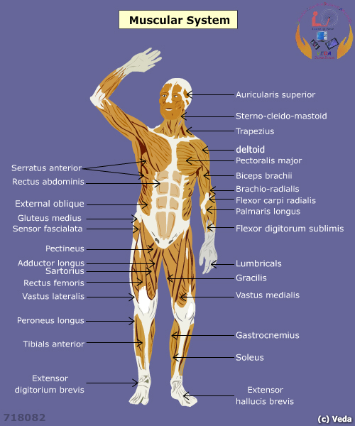
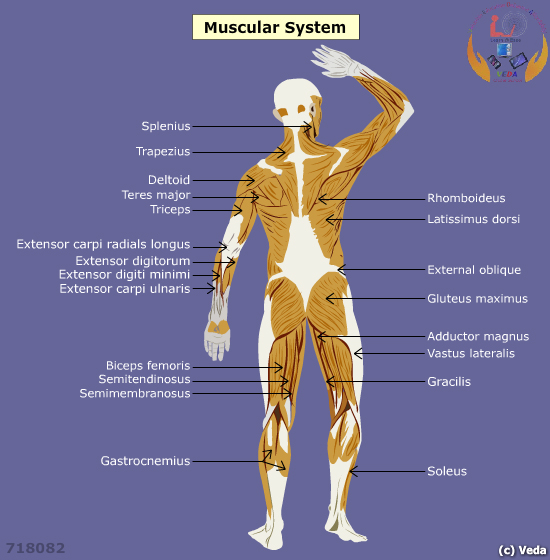
Skeletal muscleSkeletal muscle contraction is voluntary, and is therefore under the control of the somatic nervous system. Striated muscle tissue composes the skeletal muscle. It appears striated due repetitive arrangements of sarcomere units. Skeletal muscle fibers are tubular, striated, and are comprised of long multinucleated cells. A whole muscle contains bundles of muscle fibers termed fascicles and a layer of connective tissue surrounds each fascicle. Endomysium surrounds each muscle fiber and perimysium helps to bind muscle fibers into bundles termed as fascicles. These bundles then group together to form muscle, enclosed in a sheath of epimysium. Skeletal muscles attach to bones through a dense connective tissue called tendons.
Skeletal muscles fibers are composed of three types: fast twitch (II), slow twitch (I) and intermediate. Fast twitch fibers have a large diameter and depend on anaerobic metabolism. Slow twitch fibers are smaller in diameter, contain high concentrations of myoglobin (gives red color), and contain numerous mitochondria in order to perform aerobic respiration. Intermediate fibers resemble fast twitch because they contain little myoglobin but resemble slow twitch because they contain more mitochondria than fast twitch. They are also called Type IIa fibers. The ratios of the three types of fibers are determined genetically. Slow fibers are referred to as red muscle whereas fast fibers are often called white muscle.


