Learning Objectives
- Describe the functions of bone
- Contrast the structure of bones found in the axial and appendicular skeleton
- Identify the parts and locations of the major bones present in human body
- Compare mechanisms for bone ossification
- Describe the mechanism for fracture repair
- Describe disorders associated with the skeletal system
Introduction
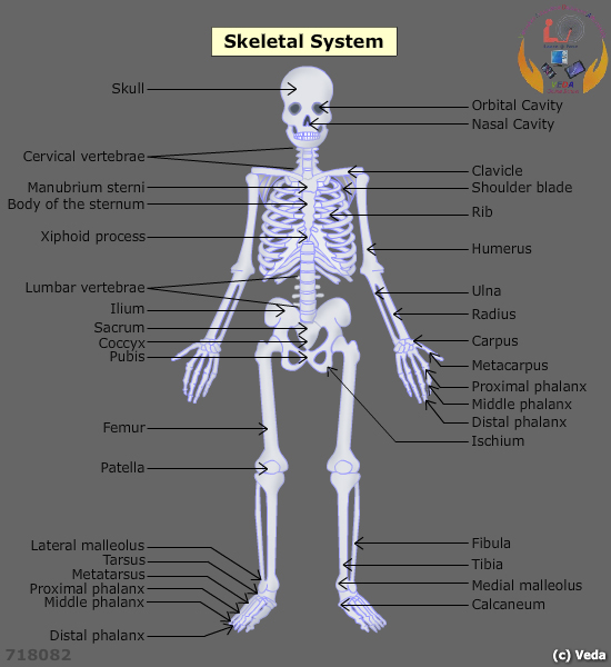
The skeletal system provides and protection support in living organisms. In the human body, the skeletal system consists of 206 bones, which work in coordination with muscles to enable movement. The skeletal system consists of two branches called the axial and appendicular skeletons, each of which is further divided into subsections.

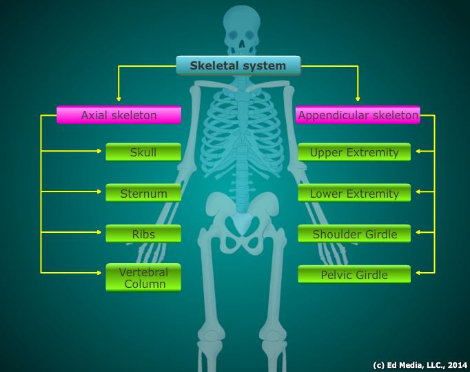
Axial skeletalThe axial skeleton consists of bones that form the axis of the body, providing support and protection to the organs of the head, neck, and trunk.
- The skull: the bony framework of the head, consisting of eight cranial bones.
- Cranial Bones: serve as a protective framework of bones around the brain.
- Frontal Bones: form part of forehead, cranial cavity, brow ridges, and nasal cavity
- Parietal Bones: the left and right parietal bones form the superior and inferior portions of the cranium.
- Temporal Bones: the right and left temporal bones form the lateral walls of cranium. They also house the external ear.
- Occipital Bones: form the posterior and inferior portions of the cranium, and are attached to the neck muscles to provide articulation to the neck.
- Sphenoid Bones: help form the floor of the cranium and the eyes orbit.
- Ethmoid Bones: form the roof of the nasal cavity as well as the medial portions of the orbits.
- Sutures: the immovable joints between the bones of the skull.
- Sagittal suture: connect the parietal bones.
- Coronal suture: meeting point of parietal bones and the frontal bones. Lambdoidal suture: meeting point of the parietal and occipital bones.
- Squamous suture: meeting point of the parietal and temporal bones.
- Facial Bones: help make up the upper and lower jaw as well other facial structures.
- Mandible: the lower jawbone, which helps to form the free joint in the head which can rotate in any direction. The mandible enables chewing action by articulating with the temporal bones at the temporomandibular joints
- Maxilla: the upper jawbones, which form part of the nose, orbits, as well the roof of the mouth.
- Palatine: the left and right palatines form a portion of the nasal cavity and posterior portion of mouth roof.
- Zygomatic: the left and right zygomatic bones form the cheekbones and a portion of orbit.
- Nasal: the left and right nasal bones form the superior portion of the bridge of the nose.
- Lacrimal: the left and right lacrimal bones form the orbits.
- Vomer: form a part of nasal septum and also helps divide the nostrils.
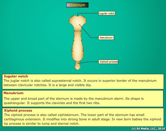
- The sternum: a flat, dagger-shaped bone found in middle of the chest that connects the ribs to for the ribcage, which provides protection to the heart, lungs, and major blood vessels. The sternum is composed of the manubrium, body, and xiphoid process. If the sternum were not present, there would be a large hole though the middle of the chest. The sternum thus protects the major organs of the chest.
- Manubrium: top portion of the sternum that is also called the handle. It is connected to the first two ribs.
- Body: middle portion of that is also called the blade or the gladiolus. The body helps connect the third along with seventh ribs directly. It indirectly connects eighth through tenth ribs.
- Xiphoid process: Bottom of the sternum is formed with the help of xiphoid process. It is also called tip.
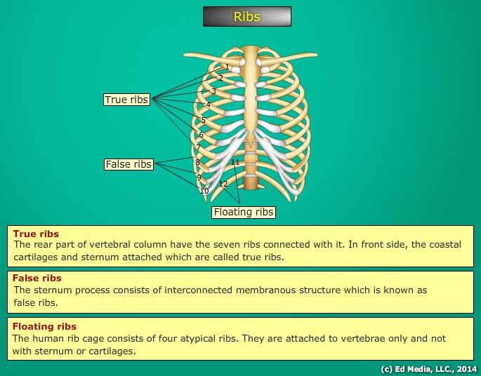
- The Ribs: flat, thin, curved bones that form a protective cage over the organs in the upper portion of the body. Twenty-four bones arranged in 12 pairs form the ribs. These bones are divided into three categories.
- True Ribs: the first seven bones in the ribs. Spines are connected with true ribs at back. Costal cartilage helps to connect the true ribs directly with breastbone or sternum.
- False Ribs: the next three bones following the true ribs. These ribs, compared to true ribs, are shorter and are connected in the back with the help of spine. False ribs are attached to the lowest true rib instead of connecting to the sternum directly.
- Floating Ribs: the remaining two sets of rib bones are called floating ribs. Floating ribs are considered the smallest ribs.
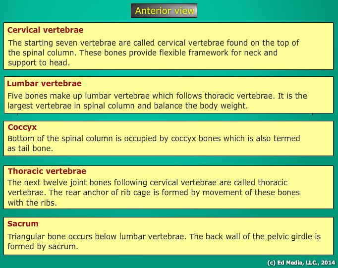
- Vertebral Column: comprised of 33 pairs of irregularly shaped bones called vertebrae. It is also called the backbone, spinal column, or spine. Based on the location of the backbone, the 33 vertebras are divided into five categories.
- Cervical vertebrae: the first seven vertebrae found on top of the spinal column. These bones provide a flexible framework for the neck as well support to the head. The first cervical vertebrae are named “atlas,” followed by the “axis”, the second vertebrae. The shape of the atlas helps the head to nod “yes” and the shape of the axis allows the head to shake “no.”
- Thoracic vertebrae: the next twelve vertebrae following the cervical vertebrae. Movement of these bones with the ribs forms the rear anchor of the ribcage. Thoracic vertebrae increase in size from top to bottom and are comparatively larger than cervical vertebrae.
- Lumbar vertebrae: comprised of five bones that come after the thoracic vertebrae. It is the largest vertebrae in the spinal column. The lumbar vertebrae balance and support most of the body weight. It is also attached to many of the back muscles.
- Sacrum: the triangular bone found just below the lumbar vertebrae. The back wall of the pelvic girdle is formed by sacrum.
- Coccyx: coccyx bones occupy the bottom of the spinal column, which is also called the tailbone.
Appendicular SkeletonBones that comprise the appendicular skeleton anchor the appendages to the axial skeleton.
- The upper Extremities: the arm, the forearm, and the hand comprise the upper extremity.
(i) The Arm – also called brachium, the arm is the region found between the shoulder and the elbow. The humerus is the long bone of the arm and is the longest bone of the upper extremity. The head, or top, of brachium is large, smooth, and rounded, fitting into the scapula in the shoulder. Two depressions are found at the bottom of humerus. The humerus is connected to the ulna and radius of the forearm at the depression. Radius and ulna together make up the elbow.
(ii) The Forearm: the region between the elbow and the wrist. The radius on the lateral side and ulna on the medial side form the forearm when viewed in the anatomical position. The radius, rather than the ulna, enables movement of the. The top of each bone connects to the humerus of the arm and the bottom of each connects to the bones of the hand.
(iii) The Hand: comprised of 27 bones and three parts–wrist, palm, and five fingers.
- Wrist: also called the carpus, the wrist is made up of 8 carpal bones. Carpal bones are bound together by ligaments. These bones in turn are arranged into two rows of four each. The top row of bones (the row closest to the forearm) from the lateral (thumb) side to the medial side holds the scaphoid, lunate, triquetral, and pisiform bones. Trapezium, trapezoid, capitates, and hamate form the second row from lateral to medial sides.
- Metacarpals: also known as the palm, comprised of five metacarpal bones, one aligned with each of the fingers. The metacarpals are numbered from I to V starting with the thumb. Wrist bones are connected at the base of metacarpal bones and the bones of fingers are connected to the heads. The heads of metacarpals form the knuckles of a clenched fist.
- Phalanges: bones making up the fingers. They are composed of 14 bones. A single finger bone is termed as phalanx and is arranged in three rows. The first row is termed as proximal row, which is the closest row to the metacarpals. The second row is called the middle row, followed by the distal row and is considered the farthest row. Similar to carpals, phalanges are numbered from I to V starting from the thumb. Each finger is provided with proximal phalanx, a middle phalanx and a distal phalanx, except the thumb, which does not contain a middle phalanx.
- The Lower Extremities: comprised of the bones of the thigh, leg, foot and patella.
- The Thigh: the region between the hip and the knee. It is comprised of a single bone termed the femur or thighbone. It is the longest, strongest, and largest bone in the human body.
- The leg: the region between the knee and the ankle. The fibula and the tibia form the leg. The fibula is arranged facing away from the bone and the tibia (shin bone) is arranged on the side nearest to the medial side of the body. Connecting tibia to the femur forms the knee joint. The talus is a foot bone that rests on top of the calcaneus and is connected to tibia. Navicular bone is found in front of talus. Bones emerging out from medial to lateral sides are the medial, intermediate, lateral cuneiform bones, and cuboid bone.
- Metatarsals and phalanges: bones of foot similar to bones of hand and are termed as metatarsals and phalanges. The metatarsals and phalanges of the foot are numbered I to V starting with the big toe. In order to support the body’s weight, the first metatarsal bone is larger than the others. The 14 phalanges of the foot, as with the hand, are formed in a proximal, middle and a distal row. It is also attached with the big toe or hallux, which has only a proximal and distal phalanx.
- The Patella: also called the kneecap, which is a large triangular sesamoid bone found between the femur and the tibia. It is formed in response to the strain in the tendon forming the knee. The patella protects the knee joint. The bones of the lower extremities are considered the heaviest, largest, and strongest bones in the body, as they are able to bear the entire weight of the body.
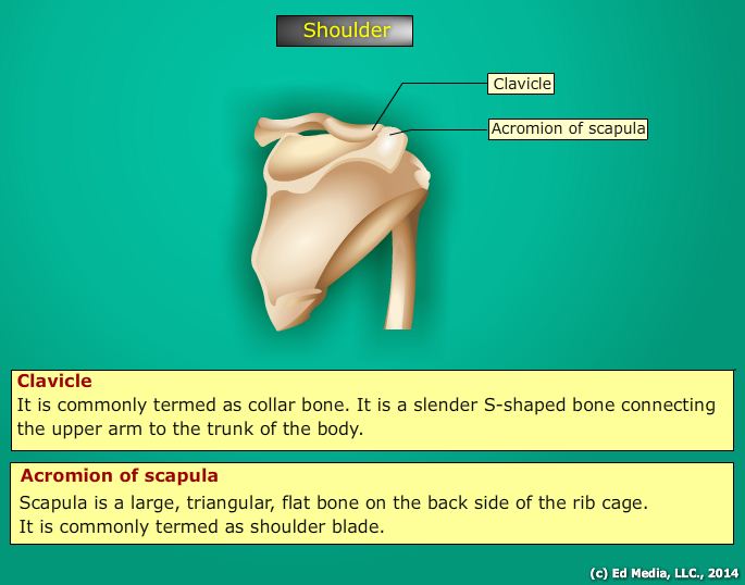
- The shoulder and pelvic girdle: the shoulder girdle is also known as the pectoral girdle. It is an incomplete ring composed of four bones, where two clavicles and two scapulae meet. The primary function of the shoulder girdle is to provide attachment points for numerous muscles that allow the shoulder and elbow joints to move. It enhances the joint between the upper extremities (the arms) and the axial skeleton.
- Clavicle: commonly known as the collarbone. It is a slender S-shaped bone connecting the upper arm to the trunk of the body. The clavicle helps to hold the shoulder joint away from the body in order to allow freedom in movement. The sternum is connected to the clavicle at one end and the scapula is connected to the other end.
- Scapula: a large, triangular, flat bone on the backside of the rib cage. It is commonly known as the shoulder blade. The scapula serves as an attachment for several muscles as the second rib is over layered through seventh rib. The glenoid cavity is a shallow depression found on the scapula that serves as head of the humerus to fit in.
- Pelvic Girdle: also termed as hip girdles. It serves several important functions in body. It is composed of two coxal (hip) bones, also known as ossa coxae. The ilium, ischium and pubis are the three points forming the coxal bones during childhood. The three coxal bones are fused together to form a single bone in adults. The two coxal bones meet on either side of the sacrum at the back. The weight of the body from vertebral column is supported by pelvic girdle. It protects the lower organs are such as urinary bladder, reproductive organs, and the developing fetus of pregnant woman. The 2 hipbones, the sacrum and the coccyx form the pelvis. Female pelvis is larger and wider than the male pelvis that is taller and narrower.


